Posted in Caregiving

How Alzheimer’s disease spreads throughout the brain

File 20180105 26154 13d7znc.png?ixlib=rb 1.1
Harmful tau protein spreads through networks.
Author provided

Thomas E Cope, University of Cambridge

Alzheimer’s disease is a devastating brain illness that affects an estimated 47m people worldwide. It is the most common cause of dementia in the Western world. Despite this, there are currently no treatments that are effective in curing Alzheimer’s disease or preventing its relentless progression.

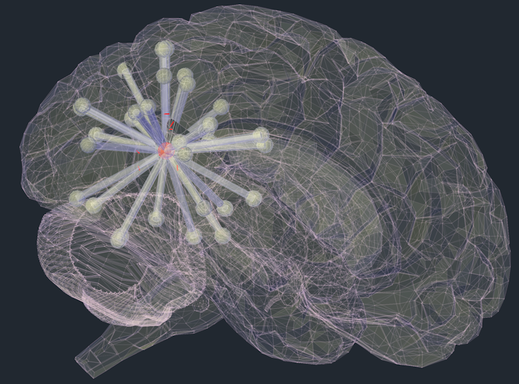
Alzheimer’s disease is caused by the build-up of two abnormal proteins, beta-amyloid and tau. Tau is particularly important because it causes neurons and their connections to die, preventing brain regions from communicating with each other normally. In the majority of cases, tau pathology first appears in the memory centres of the brain, known as the entorhinal cortex and hippocampal formation. This has been shown to occur many years before patients have any symptoms of disease.

Over time, tau begins to appear in increasing quantities throughout the brain. This causes the characteristic progression of symptoms in Alzheimer’s diseases, where initial memory loss is followed by more widespread changes in thinking and behaviour that lead to a loss of independence. How this occurs has been controversial.

Transneuronal spread

In our study, published in Brain, we provide the first evidence from humans that tau spreads between connected neurons. This is an important step, because stopping this spread at an early stage might prevent or freeze the symptoms of Alzheimer’s disease.

This idea, called “transneuronal spread”, has been proposed before and is supported by studies in mice. If abnormal tau is injected into a healthy mouse brain, it quickly spreads and causes the mice to manifest dementia symptoms. However, it had not previously been shown that this same process occurs in humans. The evidence from mouse studies was controversial, as the amount of tau injected was relatively high, and disease progression occurred much more rapidly than it does in humans.

Artist’s impression of tau spreading between connected neurons.
Author provided

In our study, we combined two advanced brain imaging techniques. The first, positron emission tomography (PET), allows us to scan the brain for the presence of specific molecules. With this, we were able to directly observe the abnormal tau in living patients, to see exactly how much of it was present in each part of the brain.

The second, functional magnetic resonance imaging (fMRI), measures blood flow in the brain in real time. This allowed us to observe the activity produced by brain regions communicating with each other. For the first time, by scanning the same people with both methods, we were able to directly relate the connections of the brain to the distribution of abnormal tau in living humans with Alzheimer’s disease.

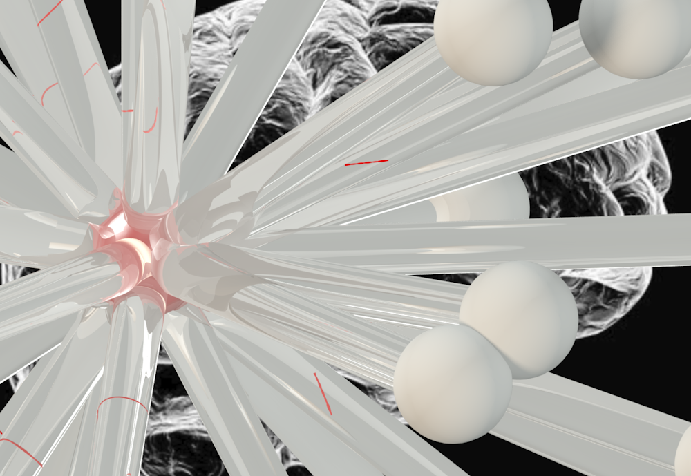
We used a mathematical technique called “graph analysis” to analyse brain connectivity. This technique involved splitting the brain up into 598 regions of equal size. We then treated the connectivity between regions like a social network, assessing factors such as the number of contacts a brain region had, how many “friendship” groups it took part in, and how many of a brain region’s contacts were also contacts of each other.

In a flu epidemic, people with a large number of social contacts are most likely to become infected and then to pass the infection on to others. Similarly, the transneuronal spread hypothesis predicts that strongly connected brain regions will accrue most tau. This is what we observed. This relationship was present within each brain network individually, as well as across the whole brain.

We were also able to exclude potential alternative explanations for the appearance of tau throughout the brain. It had previously been suggested that tau might appear at brain regions that were vulnerable because of high metabolic demand or a lack of support from their neighbours. While it is possible that these factors are important in neuronal death, our observations were not consistent with them being the primary drivers of the initial accumulation of abnormal tau.

In addition, by looking at patients with a range of disease severity, from mild cognitive impairment through to established Alzheimer’s disease, we were able to disentangle the causes of tau accumulation from its consequences. We showed that increasing amounts of tau in Alzheimer’s disease caused the brain to become less connected overall, and the connections that remained became increasingly random.

Long-range connections

Finally, we contrasted the findings in Alzheimer’s disease to a rarer condition called progressive supranuclear palsy (PSP), which affects approximately three in every 100,000 people. This condition is also caused by tau, but it remains confined to the base of the brain. We demonstrated that in PSP the evidence did not support transneuronal spread. This might be because of the different structure of abnormal tau pathology in the two diseases. In Alzheimer’s disease, tau is present in “paired helical filaments”, while in PSP it is in “straight filaments”.

Damaged communications.
Shutterstock

We showed that as PSP progresses, direct long-range connections are preferentially damaged, meaning that information had to take a more indirect route across the brain. This might explain why, when asked a question, patients with PSP usually respond slowly but correctly.

The ConversationOverall, evidence of transneuronal spread in humans with Alzheimer’s disease provides proof of concept for exciting new treatment strategies to lock up tau pathology before it can cause significant damage.

Thomas E Cope, Academic Clinical Fellow, University of Cambridge

This article was originally published on The Conversation. Read the original article.

Posted in Caregiving, International Policies, Research & Best Practice

The onset of Alzheimer’s disease: the importance of family history

News Release
February 26, 2018 | QUEBEC – The onset of Alzheimer’s disease: the importance of family history

You’re about to turn 60, and you’re fretting. Your mother has had Alzheimer’s disease since the age of 65. At what age will the disease strike you? A Canadian study published in JAMA Neurology shows that the closer a person gets to the age at which their parent exhibited the first signs of Alzheimer’s, the more likely they are to have amyloid plaques, the cause of the cognitive decline associated with the disease, in their brain.

In this study involving a cohort of 101 individuals, researcher Sylvia Villeneuve (Douglas Mental Health University Institute; CIUSSS de l’Ouest-de-l’Île-de-Montréal) shows that the difference between a person’s age and the age of their parent at the onset of the disease is a more important risk factor than their actual age.

A 60-year-old whose mother developed Alzheimer’s at age 63 would be more likely to have amyloid plaques in their brain than a 70-year-old whose mother developed the disease at age 85,” explains Villeneuve, an assistant professor at McGill University and a core faculty member at The Neuro’s McConnell Brain Imaging Centre.

Her team of scientists also found that the genetic impact of Alzheimer’s disease is much greater than previously thought.

“Upon examining changes in the amyloid biomarker in the cerebrospinal fluid samples from our subjects, we noticed that this link between parental age and amyloid deposits is stronger in women than in men. The link is also stronger in carriers of the ApoE4 gene, the so-called ‘Alzheimer’s gene’,” says Villeneuve.

Towards earlier detection of the disease

The researcher and her team successfully duplicated their results in two independent groups, one, consisting of 128 individuals from a University of Washington-St. Louis cohort, the other consisting of 135 individuals from a University of Wisconsin-Madison cohort. They also reproduced their results using an imaging technique that enables one to see amyloid plaques directly in the brains of living persons.

Their study is paving the way for the development of inexpensive methods for the early identification of people at risk for Alzheimer’s disease. According to the Alzheimer Society of Canada, 564,000 Canadians currently have Alzheimer’s disease or another form of dementia. The figure will be 937,000 within 15 years. Presently, there is no truly effective treatment for this disease.

This research was funded by grants from a Canadian research chair, the Canadian Institutes of Health Research, the Canadian Foundation for Innovation, the Canadian Brain Research Fund, the Alzheimer Society of Canada, and the Fonds de recherche du Québec — Santé.

The article entitled “Proximity to parental symptom onset and amyloid burden in sporadic Alzheimer’s disease” was published in JAMA Neurology on February 26, 2018. DOI:10.1001/jamaneurol.2017.5135

Source: http://www.douglas.qc.ca/?locale=en

Posted in Ageing & Culture, Caregiving, Therapeutic Activities

Just the two of us: Holding hands can ease pain, sync brainwaves

News Release
Februarypexels-photo-325884.jpeg 28, 2018 | COLORADO – Just the two of us: Holding hands can ease pain, sync brainwaves

Reach for the hand of a loved one in pain and not only will your breathing and heart rate synchronize with theirs, your brain wave patterns will couple up too, according to a new study.

The study, by researchers with CU Boulder and University of Haifa and published in the journal Proceedings of the National Academy of Sciences (PNAS) this week, also found that the more empathy a comforting partner feels for a partner in pain, the more their brainwaves fall into sync. And the more those brain waves sync, the more the pain goes away.

Key takeaways
  • Holding the hand of a loved one in pain can synchronize breathing, heart rate and brain wave patterns.
  • The more empathy a comforting partner feels for a partner in pain, the more their brainwaves fall into sync.
  • Increased brain synchronization is associated with less pain.

“We have developed a lot of ways to communicate in the modern world and we have fewer physical interactions,” said lead author Pavel Goldstein, a postdoctoral pain researcher in the Cognitive and Affective Neuroscience Lab at CU Boulder. “This paper illustrates the power and importance of human touch.”

The study is the latest in a growing body of research exploring a phenomenon known as “interpersonal synchronization,” in which people physiologically mirror the people they are with. It is the first to look at brain wave synchronization in the context of pain, and offers new insight into the role brain-to-brain coupling may play in touch-induced analgesia, or healing touch.

Goldstein came up with the experiment after, during the delivery of his daughter, he discovered that when he held his wife’s hand, it eased her pain.

“I wanted to test it out in the lab: Can one really decrease pain with touch, and if so, how?”

He and his colleagues at University of Haifa recruited 22 heterosexual couples, age 23 to 32 who had been together for at least one year and put them through several two-minute scenarios as electroencephalography (EEG) caps measured their brainwave activity. The scenarios included sitting together not touching; sitting together holding hands; and sitting in separate rooms. Then they repeated the scenarios as the woman was subjected to mild heat pain on her arm.

Merely being in each other’s presence, with or without touch, was associated with some brain wave synchronicity in the alpha mu band, a wavelength associated with focused attention. If they held hands while she was in pain, the coupling increased the most.

Researchers also found that when she was in pain and he couldn’t touch her, the coupling of their brain waves diminished. This matched the findings from a previously published paper from the same experiment which found that heart rate and respiratory synchronization disappeared when the male study participant couldn’t hold her hand to ease her pain.

“It appears that pain totally interrupts this interpersonal synchronization between couples and touch brings it back,” says Goldstein.

Subsequent tests of the male partner’s level of empathy revealed that the more empathetic he was to her pain the more their brain activity synced. The more synchronized their brains, the more her pain subsided.

How exactly could coupling of brain activity with an empathetic partner kill pain? More studies are needed to find out, stressed Goldstein. But he and his co-authors offer a few possible explanations. Empathetic touch can make a person feel understood, which in turn – according to previous studies – could activate pain-killing reward mechanisms in the brain.

“Interpersonal touch may blur the borders between self and other,” the researchers wrote.

The study did not explore whether the same effect would occur with same-sex couples, or what happens in other kinds of relationships. The takeaway for now, Pavel said: Don’t underestimate the power of a hand-hold.

“You may express empathy for a partner’s pain, but without touch it may not be fully communicated,” he said.

Irit Weissman-Fogel, of University of Haifa, and Guillaume Dumas and Simone Shamay-Tsoory, of Florida Atlantic University, contributed to this study. It was supported with a grant from the Binational Science Foundation.

Source: https://www.colorado.edu/today/2018/02/28/just-two-us-holding-hands-can-ease-pain-sync-brainwaves?utm_source=colorado.edu&utm_medium=Hold%20hands%20to%20ease%20a%20lover%27s%20pain%2C%20and%20your%20brains%20couple%20up%2C%20too&utm_campaign=Homepage&utm_

Written by Lisa Ann Marshall.

Posted in Caregiving, Research & Best Practice, The Built Environment

Learning from zoos – how our environment can influence our health

File 20170517 24330 1j3hzlq.jpg?ixlib=rb 1.1

CoolR/Shutterstock

Emmanuel Tsekleves, Lancaster University

We are told that we are a nation of couch potatoes, lacking the will and the strength to turn around the obesity tanker. We all need a little help in our quest for a healthier life and design can play a crucial part. If we designed our towns, cities, homes and workplaces more like animal experts design zoos, we could be one step nearer to reaching our fitness goals – as long as we can have some fun along the way.

It is reported that British people will be the fattest in Europe by 2025 and that if we want to reverse this we should have a healthier lifestyle by exercising more and eating less. But we are often made to feel guilty for not sticking to theses healthy lifestyle plans. I would suggest that before we start blaming people for adopting sedentary lifestyles, we should be taking a step back to look at the design of the environments, towns and cities in which we live.

The link between the design of the built and natural environment and its role in our health and well-being has been well explored. Now new research, led by Lancaster University, on “design for health” suggests that the environment, including buildings, cities, urban spaces and transport infrastructure, is closely linked to the lifestyles we adopt.

What is abundantly clear is that, as we shape our environment, it is also shaping us. Our psychological, physiological and physical status as well as our interactions with other people and with the natural environment are all affected. A key challenge that governments and policy makers worldwide are facing is how our built environment and infrastructure should be shaped to support healthier behaviours to prevent disease.

First, we should stop focusing on methods that tell people what to (or not to) do and which attempt to change their behaviour simply through media campaigns and punitive measures, such as tax schemes. While seeking to minimise the barriers that prevent healthy behaviours, we should make sure that the design of new environments is taken into account.

Looking to zoos

A good model would be to look at how zoos are designed. Before a zoo is built, it is common practice for zoologists, biologists, animal psychologists, nutritionists, architects, designers and landscape architects to work closely together to create an environment that optimises the living conditions for the animals.

Important environmental elements, such as vegetation, habitat, lighting, materials and each animal’s requirements are taken into account. The ultimate aim is to design an environment that fully supports the animals’ physical, psychological and social well-being. Ironically, we do not seem to make the same demands when a town, neighbourhood or workplace environment for humans is planned and designed.

Another opportunity that has recently emerged is the healthy new town NHS initiative. The aim is to radically rethink how we live and take an ambitious look at improving health through the built environment. Ten demonstrator towns will be built across England with community health and well-being as their main focus. Clinicians, designers and technology experts will reimagine how healthcare can be delivered in these places. Although this is a step in the right direction, what it is currently missing is the more holistic approach we have seen in the design of the zoos.

A crucial element in designing these towns so they are places that people would want to live in, is to include community members in their creation. This strategy would help design-in health-promoting behaviours, such as access to healthy food outlets or green spaces in which people can walk and exercise.

Embracing playfulness

Playful design – the mapping of playful experiences from games and toys to other non-game contexts – can play an important role here in inviting and encouraging people towards healthier alternatives. For example, the piano stairs project in Stockholm, which converts the metro stairs into a giant functioning piano keyboard – much like the piano made famous in the Tom Hanks movie Big (1988) – demonstrates great promise. It encourages commuters to opt for the intriguing new stairway instead of the escalators to enjoy making musical movements as they go up and down.

A project in The Netherlands, meanwhile, illustrates how everyday street furniture, such as lampposts, benches and bollards, can be inexpensively converted into impromptu exercise devices, inviting people to engage in casual activity and socialise with their neighbours. We could therefore envisage several other contexts were playfulness can transform mundane everyday activities into fun ones that encourage people into a more active and social lifestyle.

We could convert building walls into activity walls to encourage stretching of arms and legs through touch; redesign public squares and walkways into interactive dance floors that invite movement and guide you through a city; and transform workplace spaces and public places into “playgrounds” that boost movement and productivity and decrease lethargy.

The Conversation

So there you have it. If we want to be a nation of lean, mean and healthy citizens we need to learn from zoos and the animals that live in them. And we need to embrace playfulness and enjoy the place where we live. That way, we can tackle life with a hop, skip and a jump.

Emmanuel Tsekleves, Senior Lecturer in Design Interactions, Lancaster University

This article was originally published on The Conversation. Read the original article.

Posted in Caregiving, International Campaigns, International Policies, Therapeutic Activities

Bilingualism could offset brain changes in Alzheimer’s

A Concordia study sheds light on how language history relates to brain plasticity

News Release
February 6, 2018 | QUEBEC – Bilingualism could offset brain changes in Alzheimer’s

After more than a decade of research, this much we know: it’s good for your brain to know another language.

A new Concordia study goes further, however, focusing specifically on the effects of knowing a second language for patients with Alzheimer’s disease (AD) and mild cognitive impairment (MCI; a risk state for AD).

“Most of the previous research on brain structure was conducted with healthy younger or older adults,” says Natalie Phillips, a professor in the Department of Psychology.

“Our new study contributes to the hypothesis that having two languages exercises specific brain regions and can increase cortical thickness and grey matter density. And it extends these findings by demonstrating that these structural differences can be seen in the brains of multilingual AD and MCI patients.”

Phillips’s study, led by recent Concordia psychology grad Hilary D. Duncan (PhD 17), is soon to be published in Neuropsychologia(Jan, 2018).

New methods: Enter the MRI

Phillips and her team are the first to use high-resolution, whole-brain MRI data and sophisticated analysis techniques to measure cortical thickness and tissue density within specific brain areas.

Namely, they investigated language and cognition control areas in the frontal regions of the brain, and medial temporal lobe structures that are important for memory and are brain areas known to atrophy in MCI and AD patients.

“Previous studies used CT scans, which are a much less sensitive measure,” says Phillips, founding director of Concordia’s Cognition, Aging and Psychophysiology (CAP) Lab.

The study looked at MRIs from participating patients from the Jewish General Hospital Memory Clinic in Montreal.

Their sample included 34 monolingual MCI patients, 34 multilingual MCI patients, 13 monolingual AD patients and 13 multilingual AD patients.

Phillips believes their study is the first to assess the structure of MCI and AD patients’ language and cognition control regions. It is also the first to demonstrate an association between those regions of the brain and memory function in these groups, and the first to control for immigration status in these groups.

“Our results contribute to research that indicates that speaking more than one language is one of a number of lifestyle factors that contributes to cognitive reserve,” Phillips says.

“They support the notion that multilingualism and its associated cognitive and sociocultural benefits are associated with brain plasticity.”

What’s next?

Phillips and her team are already building on their findings.

“Our study seems to suggest that multilingual people are able to compensate for AD-related tissue loss by accessing alternative networks or other brain regions for memory processing. We’re actively investigating that hypothesis now.”

Read the cited study, “Structural brain differences between monolingual and multilingual patients with mild cognitive impairment and Alzheimer disease: Evidence for cognitive reserve.

Source: NEW RESEARCH: Bilingualism could offset brain changes in Alzheimer’s